

What Is Integrity™?
Integrity™ is a hybrid tendon repair scaffold composed of:
- 80% Hyaff® (esterified hyaluronic acid)
- 20% PET fibers (polyethylene terephthalate)7,8
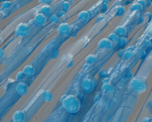
The hybrid design combines biologic support with mechanical reinforcement. The Hyaff scaffold supports cellular infiltration and tissue remodeling,4,5 while PET fibers provide durability and handling characteristics7 during implantation.
Available in multiple sizes to support shoulder, knee, hip, foot, and ankle tendon repairs.
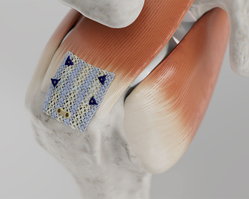
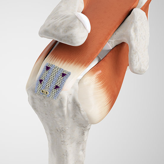 Rotator Cuff Tendon
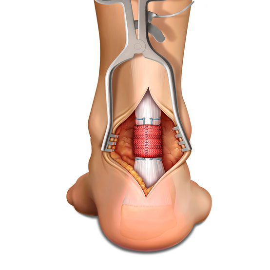 Achilles Tendon
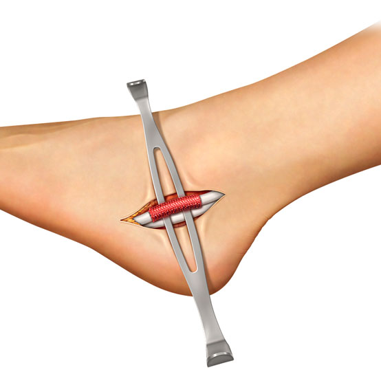 Peroneal Tendon
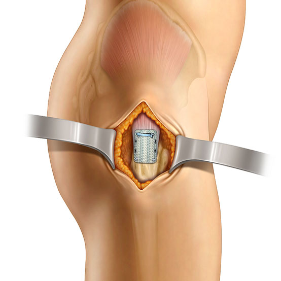 Gluteus Medius Tendon
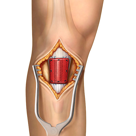 Patellar Tendon
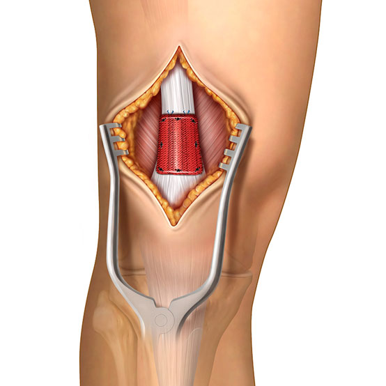 Quadriceps Tendon

Why Choose Integrity™?
Tendon augmentation supports biologic healing and tendon remodeling following repair. Integrity incorporates hyaluronic acid technology to promote cellular infiltration and tissue regeneration.
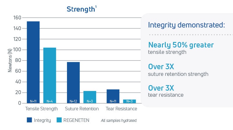
Reliable Mechanical Strength
- High tensile strength and suture retention7,8
- Maintains structural integrity when hydrated8
- Facilitates arthroscopic handling and fixation7,8
Biologic Healing Support
- HA-based scaffold promotes cell infiltration1,4,5
- Supports tissue remodeling and tendon thickening1,4,5
- Gradually resorbs as healing progresses4,5
Procedural Simplicity
- Streamlined implantation technique3
- Precise scaffold placement3
- Sterile, single-use delivery instruments3
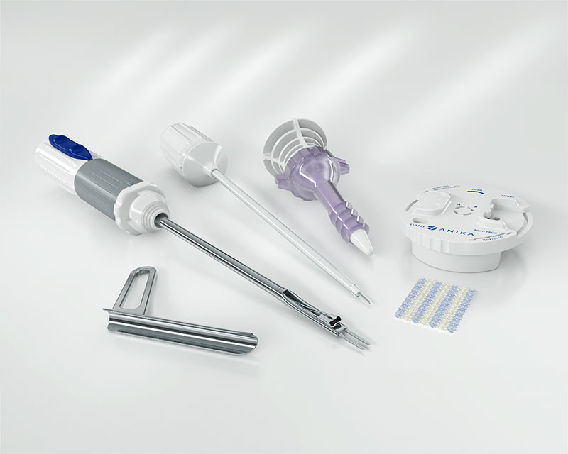

Engineered for Tendon Repair
Integrity™ combines key design features that support tendon repair:
| Feature | Clinical Relevance |
|---|---|
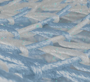
| Porous knitted architecture | Supports cellular infiltration and tissue ingrowth1 |
| Hyaff® technology | Provides a bioactive, resorbable scaffold4,5 |
| PET reinforcement | Enhances handling and mechanical strength7,8 |
| Hybrid composition | Balances biologic healing with structural support2,8 |
| Non-xenograft design | Contains no animal-derived tissue2,8 |
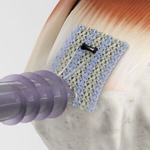
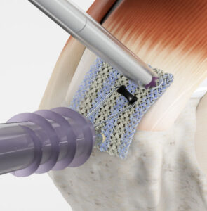
Following tendon repair, Integrity™ is positioned over the repair site as an augmentation scaffold.3 The PET component provides immediate structural support,7,8 while the Hyaff matrix promotes cellular infiltration and tissue remodeling.1,4,5 Over time, the Hyaff component gradually resorbs as new tendon tissue develops.4,5
Clinical Relevance

Evidence
In a head-to-head preclinical comparison with REGENETEN®:
- Greater fibroblast infiltration observed as early as 12 weeks1
- More organized collagen formation within the repair tissue1
- Nearly 3× greater tendon thickness at 26 weeks1
- Enhanced tissue remodeling within the resorbing scaffold1
Increased tendon thickness may contribute to a more favorable biomechanical environment and reduced tendon strain during healing.6
Clinical Highlights: Rotator Cuff Repair3
A recent white paper evaluating 29 patients treated with the Integrity Implant System for partial and full thickness rotator cuff tears revealed compelling early outcomes:
- Pain relief and functional improvement sustained over 6 months3
- No device-related complications reported3
- No evidence of retears at the 6-month follow-up3
The Integrity Implant System shows promise as a biologic strategy to support tendon healing in rotator cuff repair.3
View Full White PaperReferences
- Walsh WR, et al. Rotator Cuff Healing with Anika Technology. Data on File, Anika Therapeutics, Inc.
- Patel R, et al. Hyaluronic Acid–Biocompatible Implant System for Rotator Cuff Augmentation. Arthroscopy Techniques. 2025:103990.
- Porter DA, Pietrzak WS. The Integrity Implant System: Early Outcomes Following Rotator Cuff Repair. White Paper. Anika Therapeutics, Inc. 2025. AML-900-735 REV 01.
- Hu M, et al. Three-Dimensional Hyaluronic Acid Grafts Promote Healing and Reduce Scar Formation in Skin Incision Wounds. Journal of Biomedical Materials Research. 2003;67(1):586–592.
- Vindigni V. Hyaluronan Benzyl Ester as a Scaffold for Tissue Engineering. International Journal of Molecular Sciences. 2009;10:2972–2985. doi:10.3390/ijms10072972.
- Schlegel TF. Radiologic and Clinical Evaluation of a Bioabsorbable Collagen Implant to Treat Partial-Thickness Tears: A Prospective Multicenter Study. Journal of Shoulder and Elbow Surgery. 2017. doi:10.1016/j.jse.2017.08.023.
- Metzger A. Polyethylene Terephthalate and the Pillar™ Palatal Implant: Its Historical Usage and Durability in Medical Applications. Biomedical Engineering. 1976;11(9):301–306.
- Data on File. Anika Therapeutics, Inc.

Percutaneous Cancellous Bone Harvesting Procedure Kit
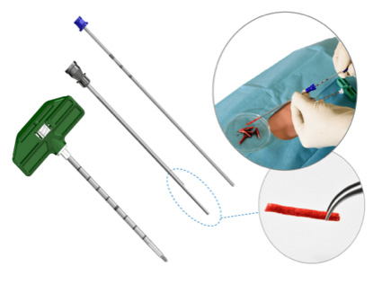
This convenience kit includes an 8 gauge bone marrow biopsy needle used for percutaneous trephine extraction of autogenous bone marrow/cancellous bone dowels. Trephine extraction of cancellous bone is considered a minimally invasive technique compared to open methods of bone extraction and has been documented to have less donor site morbidity than open procedures.1,2,3
Autograft bone is considered as the “gold standard” clinical material for grafting.4,5 Harvesting intact cancellous bone cores without disrupting the highly-organised living tissue is superior to transplanting pieces of bone. Intact grafts maintain the micro-vascular network within the graft, promoting bone callus formation/remodelling and do not exhibit extensive resorption.6,7
Intact bone exploits the biology of normal fracture healing rather than through slow creeping substitution associated with the slow incorporation of a non-vascularised graft.6 Research demonstrates the enhanced survival of a bone graft as long as its primary blood supply is preserved. A living bone graft will shorten the time for bony union because the reconstructed bone is comparable to a bone with a double fracture.6,7
The Advantages
- Graft material with osteoconductive, osteoinductive & osteogenic properties
- Graft material may be combined with autologous, allogenic, xenogenic or synthetic bone chips hydrated with high cellular marrow aspirate
- Minimally invasive technique — using an 8 Gauge Trephine Needle for bone core extraction
- Reduces donor site morbidity associated with standard harvesting techniques
Marrow Cellution Cancellous Bone Graft Harvesting
Marrow Cellution Product Information
For more information on Marrow Cellution, please download the brochure below.
↓ Download Marrow Cellution BrochureReferences
- https://www.cochranelibrary.com/cdsr/doi/10.1002/14651858.CD011783/full accessed on 13 Nov 2018.
- Missiuna PC, Gandhi HS, Farrokhyar F, et al. Anatomically safe and minimally invasive transcrestal technique for procurement of autogenous cancellous bone graft from the mid-iliac crest. Can J Surg. 2011;54(5):327–32.
- Hernigou P, et al. Morbidity of graft harvesting versus bone marrow aspiration in cell regenerative therapy. Int Orthop. 2014 Sep;38(9):1855–60.
- Block JE. The role and effectiveness of bone marrow in osseous regeneration. Med Hypotheses. 2005;65(4):740–7.
- Wang W, Yeung K. Bone grafts and biomaterials substitutes for bone defect repair. A review. Bioactive Materials. 2017;2(4):224–227.
- Bleuming SA, et al. Bone morphogenetic protein signalling suppresses tumorigenesis at gastric epithelial transition zones in mice. Cancer Research. 2007;67(17):8149–55.
- Ostrup et al. Distant transfer of a free, living bone graft by micro-vascular anastomoses. An experimental study. Plast Reconstr Surg. 1974 Sep;54(3):274–85.
Tactoset — Injectable Bone Substitute
Tactoset is a synthetic, biocompatible, hyaluronic acid-enhanced, calcium phosphate bone graft substitute material intended for permanent implantation and filling bone voids (known as bone marrow lesions) or defects of the skeletal system that are not intrinsic to the stability of bony structure.
Tactoset hardens post deployment to reinforce weaknesses created by these bone voids by converting to a poorly crystalline hydroxyapatite at body temperature. It is then resorbed and replaced by the growth of new bone during the healing process.
Components
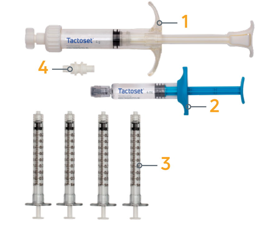
Tactoset Material Set
- Pre-filled mixing syringe containing powder
- Glass syringe containing setting solution
- Four 1 mL Luer lock syringes
- Female-female Luer lock connector
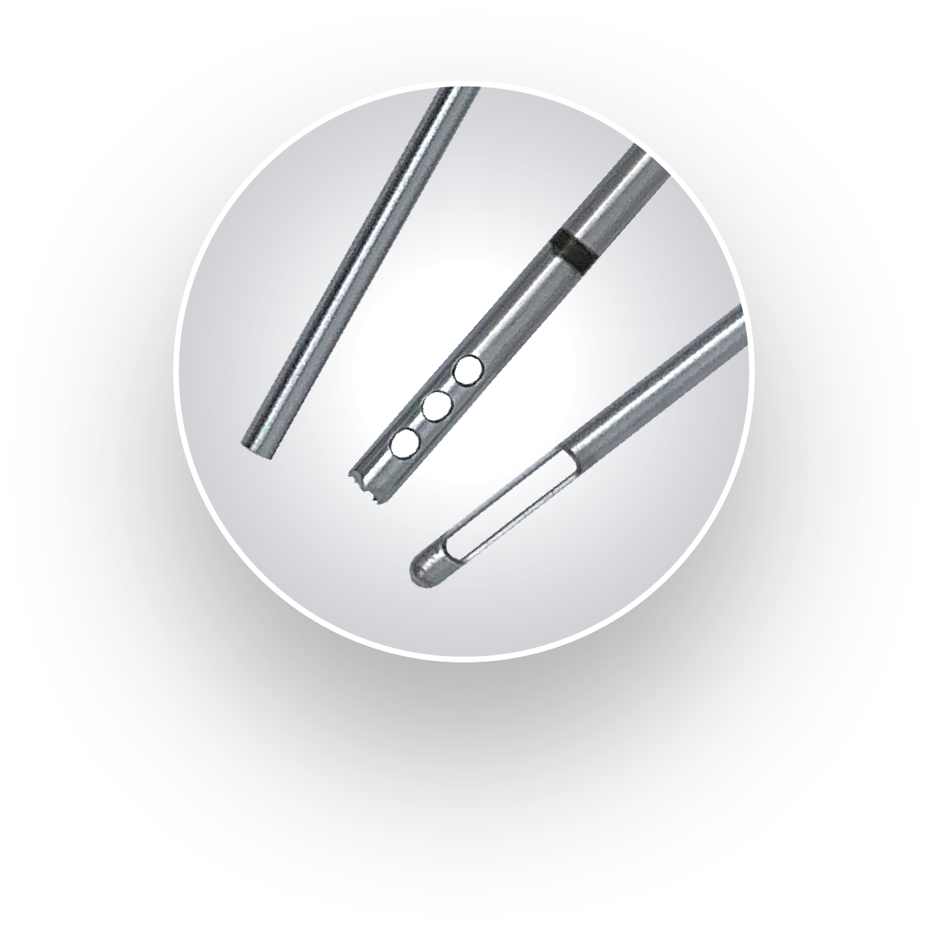
Anika Delivery Cannulas
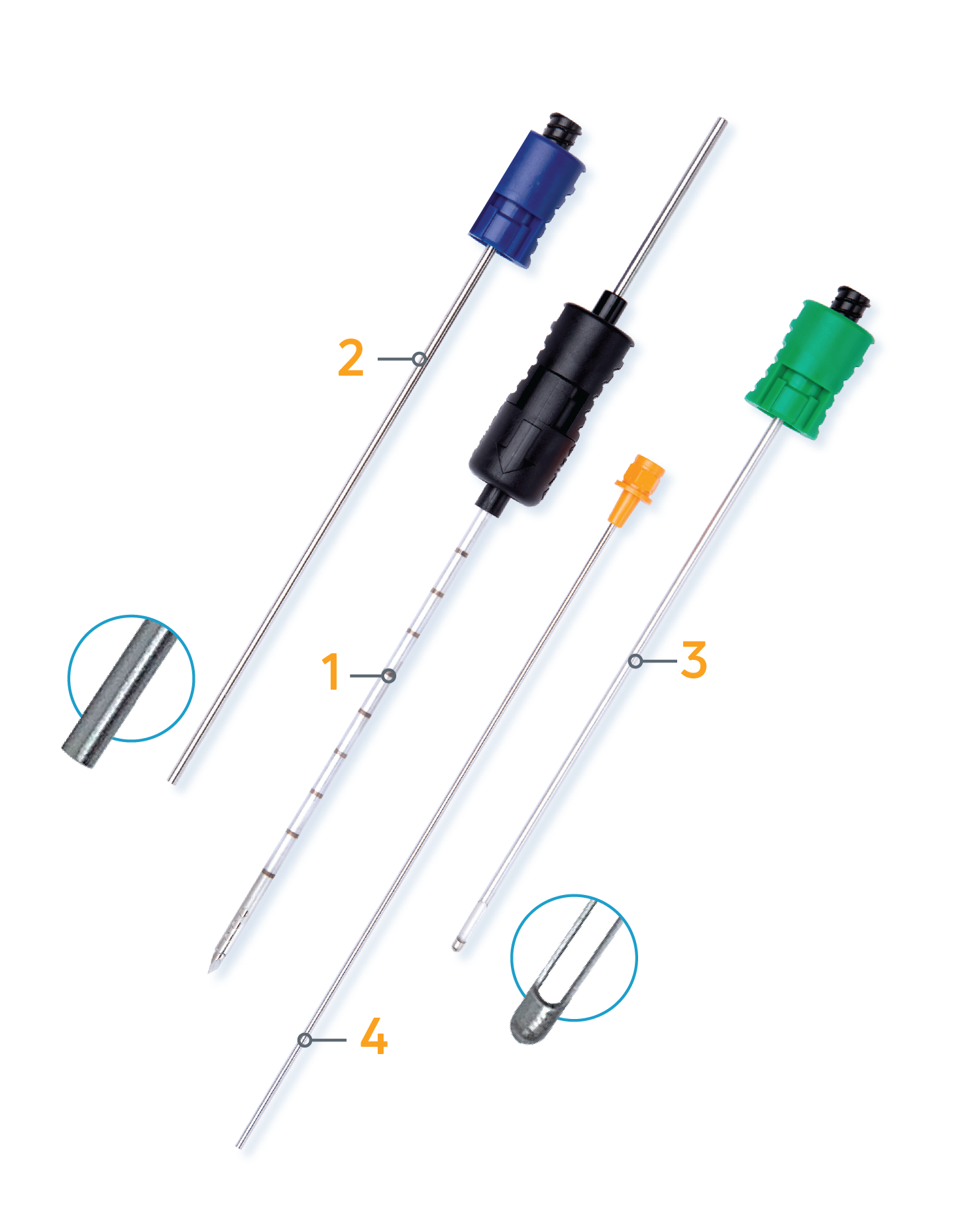
- Drillable outer cannula (with removable drillable stylet) — GAUGE: 3.05 mm (11 Ga), LENGTH: 110 mm
- Inner cannula for end delivery — GAUGE: 2.41 mm (13 Ga), LENGTH: 134.39 mm
- Inner cannula for side delivery — GAUGE: 2.41 mm (13 Ga), LENGTH: 134.39 mm
- Push rod
Anika delivery cannulas provide easier use with mini c-arm.
Therapy Advantages
Tactoset supports cell-mediated regeneration of new bone as material is resorbed.
Enhanced Flowability and Injectability
- Contains hyaluronic acid (HA), a naturally occurring substance in the human body
- HA enhances delivery of the calcium phosphate
- Highly flowable and easily injectable through minimally invasive surgery
- Provides tactile feedback to confirm filling of the bone defect
- Interdigitates into trabecular bone architecture to fill closed bone voids
Closed Mixing System
- Minimizes environmental exposure
- Enhances ease of use: no mixing bowls; no mess
Unique Cannula Design
- Allows surgeons to easily switch intraoperatively between side and end Tactoset delivery without having to remove and reinsert the outer cannula
Diagnosis
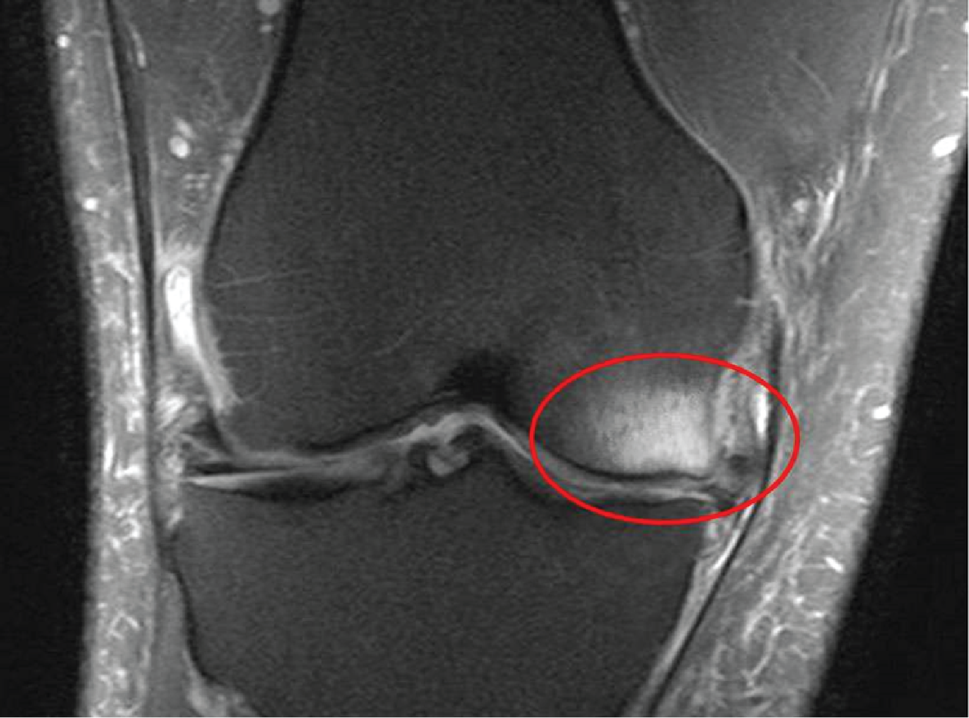
- X-rays are commonly used to determine severity of osteoarthritis, however bone marrow lesions (BMLs) cannot be detected on X-ray
- BMLs can be identified on fluid sensitive, fat suppressed magnetic resonance imaging (MRI)
- Recognised as an area of intense signal visible on T2-weighted sequences
Tactoset Product Information
For more information on Tactoset, please download the brochure below.
↓ Download Tactoset Brochure